What is a Diabetic Eye Exam?

Taking care of our eyes is crucial for maintaining overall health and well-being. Regular eye exams are pivotal in preserving good vision and detecting potential issues before they develop into complex conditions. On the other hand, comprehensive eye exams offer a more holistic and in-depth look at our eye health. A diabetic eye exam is a specialized type of comprehensive eye exam and a critical preventive care component for individuals with diabetes.
Examining the Difference in Eye Exams
Regular eye exams and comprehensive eye exams are distinct in a few different aspects. Regular eye exams focus primarily on detecting refractive errors, such as myopia or hyperopia, and evaluating the need for corrective lenses. Visual acuity, refraction, and visual field tests are standard during regular eye exams. Comprehensive eye exams include these tests but involve additional screenings offering thorough insights regarding the eye’s health — and the health of the individual. These examinations may consist of:
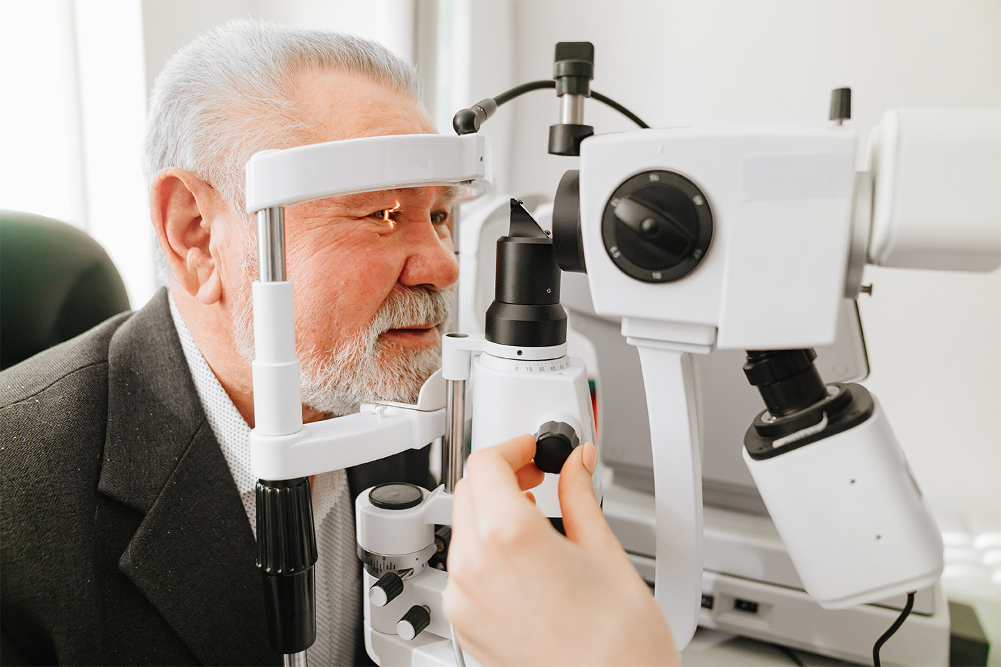
- A slit-lamp exam to determine the health of the cornea, lens, and retina;
- A tonometry test to measure intraocular pressure (a possible indicator of glaucoma when elevated);
- Pupil dilation for a more detailed view of the retina; and
- Retinal imaging to detect signs of disease or damage.
Annual comprehensive eye exams are crucial for individuals with existing eye conditions, a family history of eye diseases, or specific systemic health issues like diabetes.
The Link Between Diabetes and Eye Health
Diabetes can have a profound impact on various organs, and the eyes are no exception. High blood sugar levels associated with diabetes can lead to diabetic retinopathy. This condition damages the blood vessels in the retina and can lead to vision problems or even blindness if not addressed. Diabetes can also increase the risk of other eye problems, such as glaucoma, cataracts, or diabetic macular edema.
The Importance of Diabetic Eye Exams
Diabetic eye exams are designed to monitor and detect diabetes-related eye complications, including diabetic retinopathy. These exams consist of the same tests as a comprehensive eye exam with a specific focus on the retina. Retinal imaging methods during a diabetic eye exam may include:
- Color fundus photography: Fundus is another term for the back of the eye. A special camera takes detailed images of the retina, including the blood vessels, to identify any abnormalities and signs of health concerns.
- Optical Coherence Tomography, or OCT: This imaging provides cross-sectional views of the retina’s layers, revealing swelling or fluid leakage and allowing your provider to measure the retina’s thickness.
- Fluorescein angiography: During this test, a yellowish-colored dye called fluorescein is injected into a vein, usually in your arm. Once the dye travels to your eye, it highlights the blood vessels in your retina to help identify circulation issues.
The American Diabetes Association recommends a comprehensive eye exam annually for individuals with diabetes, even those without apparent vision problems — but it’s imperative to talk to your eye care provider about what’s right for your unique health situation. Early detection of diabetes-related eye conditions is vital to prevent vision loss.
Have Confidence in Your Eye Care
Regular eye exams for everyone, including those without diabetes, are essential for maintaining overall eye health. For individuals with diabetes, adhering to recommended eye exam schedules and communicating any vision or eye health changes to their health care provider is crucial. With decades of combined experience, our expert team of world-class doctors at Swagel Wootton Eye Institute provides the highest quality eye exams with a team-oriented approach. Contact us today to prioritize your preventive care and schedule your diabetic eye exam.


